April 14, 2021
(LOS ANGELES) – Quantum dot technology is a subject of interest for big television manufacturers, who are incorporating this technology into building their liquid crystal display (LCD) televisions. Quantum dots are tiny nanocrystals which have a special power: when exposed to light, they become luminous and give off a very pure and precise color depending on their size. The desired color can be fine-tuned by adjustment of the quantum dot’s size; using the dots in an LCD television enhances the colors on the screen and results in a better picture. Quantum dot technology can also be used in other ways, such as in solar panels and for medical uses.
One type of medical application revolves around the use of injectable hydrogels. A hydrogel is a gelatinous substance that is biocompatible with bodily tissues and has tunable elastic and mechanical properties. Some hydrogels also have shear-thinning capabilities – the ability to deform upon injection and then quickly self-recover. Injectable shear-thinning hydrogels have been used successfully for delivering therapeutic agents into the body, as well as for hemorrhage control in cases of acute bleeding or aneurysms.
For these applications, it is very important to monitor the placement of the hydrogel and its eventual degradation in the body; this requires dynamic, real-time monitoring. Quantum dots are a promising alternative for image labeling, although certain types of quantum dots are unsuitable for biomedical purposes due to their high toxicity.
A collaborative team from the Terasaki Institute for Biomedical Innovation (TIBI) has recently examined the properties of graphene quantum dots. Graphene, a form of carbon of ultra-small size, is biocompatible, stable under bodily conditions, readily available at low cost and has strong, adjustable fluorescent capabilities.
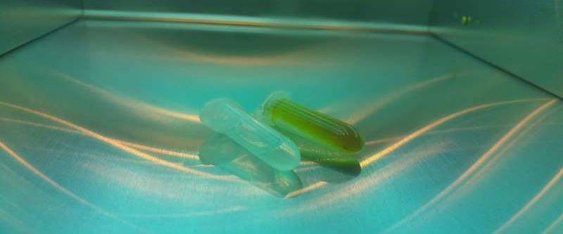
The team prepared and characterized the graphene quantum dots before mixing them with shear-thinning hydrogels. The resultant mixture was tested and optimized for its elasticity, sheer-thinning properties and biocompatibility. It was also tested for its fluorescent capabilities by using both direct observation after exposure to UV light and by imaging after injection into mouse animal models.
“After conducting our experiments, we were able to obtain the correct formulation for optimum performance of our graphene quantum dot hydrogels,” said TIBI team member Fatemeh Nasrollahi, Ph.D. “Our tests have resulted in a robust imaging method that can easily be used in the clinic.”
It is anticipated that use of these graphene shear-thinning hydrogels will offer a means of low-cost, high-quality imaging for many biomedical applications in the future.
“This work is one of many examples under our biomaterials research platform,” said Ali Khademhosseini, Ph.D., director and CEO of the Terasaki Institute for Biomedical Innovation. “The use of graphene quantum dots can provide an alternative method of medical imaging.”
Additional authors are Ali Khademhosseini, Farzana Nazir, Maryam Tavafoghi, Vahid Hosseini, Mohammad Ali Darabi, David Paramelle and Samad Ahadian.
This work was funded in part by the National Institutes of Health (HL 140951 and HL 137193).

